All Jharkhand Competitive Exam JSSC, JPSC, Current Affairs, SSC CGL, K-12, NEET-Medical (Botany+Zoology), CSIR-NET(Life Science)

SIMOTI CLASSES

Education Marks Proper Humanity

SIMOTI CLASSES

Education Marks Proper Humanity

SIMOTI CLASSES

Education Marks Proper Humanity

SIMOTI CLASSES

Education Marks Proper Humanity

SIMOTI CLASSES

Education Marks Proper Humanity

Showing posts with label CSIR-NET-LIFE-SCIENCE. Show all posts

Tuesday, June 8, 2021

Tumor Suppressor Gene p53 - CSIR NET/ DBT/ ICMR (Life Sciences)

Tumor Suppressor Gene p53

The TP53 gene codes for p53 protein (also known as TP53) of an apparent molecular mass of 53 kDa.

It is a Tumor Suppressor gene, its activity stops the formation of tumors. It is located on human chromosome 17p13.1 and encodes a polypeptide chain of 393 amino acid residues.


p53 Protein Regulation:

p53 protein is a transcription factor that regulates the expression of several genes involved in;

  • DNA repair
  • cell growth
  • apoptosis (programmed cell death)
  • antioxidant defense
  • senescence

Because mutations in the TP53 gene are found in most tumor types and are involved in the complex network of cellular events, it is described as the guardian of the genome and the cellular gatekeeper.

DNA damage and role of p53 protein
Fig: DNA damage and role of p53 protein

In a normal cell, p53 is inactivated by its negative regulator, MDM2 (mouse double minute 2). 


  • Upon DNA damage or the stresses, various pathways lead to the dissociation of the p53 and MDM2 complexes.

Once activated;

  • p53 will induce a cell cycle arrest to allow either repair and survival of the cell or apoptosis to discard the cell. For example, DNA damage activates p53.

  • In undamaged cells, p53 is highly unstable and is present at very low concentrations. This is large because it interacts with MDM2, which causes ubiquitination of p53 and targets it for proteasomal destruction. 

  • Phosphorylation of p53 after DNA damage reduces its binding to MDM2. This decreases p53 degradation, which results in a marked increase in p53 concentration in the cell.

  • In response to double-strand breaks (DSBs) in DNA, protein kinase ATM is activated, activating CHK2 kinase. 

  • ATM and CHK2 then both phosphorylate p53 at distant sites. Phosphorylation of p53 on amino acid residues in its N-terminal domain by kinases such as ATM and CHK2 alters the domain of p53. 

  • At the same time, the ATM kinase can phosphorylate MDM2 in a way that causes its functional inactivation. As a consequence of this phosphorylation of both p53 and MDM2, MDM2 fails to initiate ubiquitination of p53, p53 escapes destruction, and p53 concentrations in the cell increase rapidly. Once present in substantial amounts, p53 is then poised to evoke a series of downstream responses.

Now,

  • p53 stimulates transcription of the gene encoding a CKI protein called p21.

  • This protein binds to G1/S-CDK and S-CDK complexes and inhibits their activities, thereby helping to block entry into the cell cycle. 

  • Hence, in response to DNA damage, p53 levels increase and arrest the cell at the G1-phase of the cell cycle. 

  • If all the repairs have been made to the DNA, the cell divides normally and completes the cycle. 

  • However, if the cell still contains a mutated or duplicated DNA sequence, it dies by a suicidal apoptotic mechanism to prevent its proliferation. In those cells that have mutated or lost the p53 gene, the arrest at G1 does not occur, and the cells that have mutated genomes proliferate and become cancerous.

p53-mediated apoptosis involves the coordination of various function of p53;

  • transcription-dependent
  • transcription-independent

p53 also involves the transcription factor for numerous genes involved in apoptosis such as PUMA, NOXA.

p53 has cytosolic activities that can induce apoptosis in a transcription-independent manner. p53 can induce mitochondrially outer membrane permeabilization (MOMP), thus leading to the release of cytochrome c from the mitochondria. BCL-XL, BCL-2, and BAX may influence the voltage-dependent anion channel, which may play a key role in the regulation of cytochrome c release.


Share:

Wednesday, May 26, 2021

Gel Electrophoresis of Protein - CSIR/ DBT/ ICMR (Life Sciences)

Gel Electrophoresis of  Protein

Gel Electrophoresis:

Features:

  • Gel matrix is the stationary phase and liquid (buffer with various ions) is the mobile phase.

  • Gel matrix is made up of some polymers viz. polyacrylamide polymer or agarose polymer.

  • The gel is a three-dimensional molecular network and this molecular network has various pores through which molecules can pass. Hence, it acts as a mobile sieve.


  • The distribution of the pore size in a gel determines the size range of the ions that can be separated.

Theory of Gel Electrophoresis:

A molecule with net charge 'q' moves in an electric field with velocity 'v'.

v = E*q/f

Where, 

  • 'E' is electric field in volts/cm. 
  • 'f' is a frictional coefficient, which depends upon the shape and mass of the molecules.

Experiments are typically done at a constant voltage. Then the movement of molecules depends only on q/f.

For molecules of similar shapes, such as DNA or SDS-bound proteins, the movement depends only on the size of the molecules. Small molecules move faster than large molecules.


Methods of gel electrophoresis:

Polyacrylamide Gel Electrophoresis (PAGE):

  • Used to separate large biomolecules such as proteins and DNA.
  • It typically runs under two conditions: denaturing and native.
  • High-resolution power for biomolecules up to 1000 kDa.
  • Gel matrix has good physical stability, thus, easy to handle.
  • Sample separation is due to both molecular sieving and electrophoretic mobility.
  • No void volume in the matrix. An only continuous network of pores.
  • Small molecules move faster than larger molecules.
  • The pore size of the gel matrix is controlled by the concentration of acrylamide and N, N'-methylenebisacrylamide.
  • Caution: Monomer acrylamide is a neurotoxin and is also suspected to be a carcinogen.
acrylamide (4 to 20%)
Fig: acrylamide (4 to 20%)


N, N'-methylenebisacrylamide (1 to 5% of acrylamide)
Fig: N, N'-methylenebisacrylamide (1 to 5% of acrylamide)

Polyacrylamide Matrix
Fig: Polyacrylamide Matrix


Advantages of Polyacrylamide Gel:

  • It does not interact with protein or nucleic acid.
  • It is chemically stable, hydrophilic, free of ions.
  • It does not interfere with common staining reactions.


SDS-PAGE denaturing condition:

Discontinuous Gel Electrophoresis:

  • Two gel layers: stacking gel (upper) and resolving gel (lower).
  • These are prepared with buffers with different ionic strengths and pH.
  • Stacking gel concentrates all protein molecules into a tight band before entering the resolving gel.
  • Sodium dodecyl sulfate (SDS) is a detergent.
  • SDS binds hydrophobic regions of the denatured protein, resulting in a constant charge/mass ratio and uniform shape. Proteins are now separated by size.
  • The majority of the current is carried by the buffer ions i.e.tris, glycine, and chloride.
  • Protein ions have a negligible contribution to the current.
  • At low buffer concentration, proteins will migrate fast, resulting in smeared bands.
  • At high buffer concentration, proteins will move very slowly.
  • Visualizing the protein bands with Bromophenol blue (negatively charged and high electrophoretic mobility, runs fastest), Coomassie Brilliant Blue R-250, and Coomassie Brilliant Blue G-250.

Functions of Stacking gel:

  • The stacking gel is polymerized from a dilute acrylamide solution. The large pore size does not provide a molecular sieving effect.
  • Stacking gel ensures that all of the proteins arrive at the running gel at the same time so proteins of the same molecular weight will migrate as tight bands.

Functions of Resolving gel:

  • The resolving gel is to separate the protein based on their molecular weight.


Processes:

  • Protein purified sample incubated with Sodium Dodecyl Sulfate (SDS) to provide a uniform negative charge.
  • Sample from different animals loaded into different "wells" of an electrophoresis chamber.
  • Acrylamide gel, containing microscopic pores, separates the cathode/anode.
  • The first "well" contain loading control. A mixture of multiple proteins of known molecular mass added as a reference to experimental wells. Tracking dye added to loading control to visualize protein migration.
  • When electrical current is run through the chamber, negatively charged proteins migrate towards a positive anode.
  • Smaller proteins "slip" through resolving gel more quickly than larger bulky proteins.
  • Visualization of protein by western blotting.

SDS- Polyacrylamide Gel Electrophoresis

Fig: SDS- Polyacrylamide Gel Electrophoresis


Share:

Monday, May 17, 2021

Breast Cancer (BRCA1) gene binding partner- CSIR/ ICMR/ DBT (Life Sciences)

Breast Cancer (BRCA1) Gene Binding Partner

1.) MDC1- Gene

Protein: Mediator of DNA-damage Checkpoint protein1

Function:

  • It is required for checkpoint-mediated cell cycle arrest in response to DNA damage within both the S-phase and G2/M phases of the cell cycle.
  • It serves as a scaffold for the recruitment of DNA repair and signal-transduction proteins to discrete foci of DNA damage marked by 'Ser-139' phosphorylation of histone H2AX. 
  • It helps in phosphorylation and activation of the ATM, CHEK1, CHEK2 kinases, and stabilization of TP53 and apoptosis. ATM and CHEK2 may also be activated independently by a parallel pathway mediated by TP53BP1.

2.) BAP1

Protein: Ubiquitin carboxyl-terminal hydrolase.

Function:

  • It helps regulates the function of many proteins involved in diverse cellular processes, control cell growth and division (proliferation), and cell death.
  • It helps repair damaged DNA, control the activity of genes, and act as tumor suppressor genes.

3.) BARD1

Protein: BARC1 Associated RING Domain 1

Function:

  • E3 ubiquitin-protein ligase. 
  • BARC1-BARD1 heterodimer specifically mediates the formation of 'Lys-6'- polyubiquitin chains.
  • It coordinates a diverse cleavage range of cellular pathways such as DNA damage repair, ubiquitination, and transcriptional regulation to maintain genomic stability. 
  • It acts by mediating ubiquitin E3 ligase activity that is required for its tumor suppressor function. 
  • It forms a heterodimer with CSTF1/CSTF-50 to modulate mRNA processing and RNAP II stability by inhibiting pre-mRNA 3' cleavage.


4.) CsTF

Protein: Cleavage Stimulation Factor.

Function:

  • It is recruited by Cleavage & Polyadenylation Specificity Factor (CPSF).
  • It assembles into protein complex on the 3' end to promote the synthesis of functional polyadenine tail, which results in mature mRNA molecule ready to be exported from nucleus to cytosol for the translation process.


Share:

Sunday, May 16, 2021

Blue-White Screening Mechanism - CSIR/ ICMR/ DBT (Life Sciences)

Blue-White Screening Mechanism

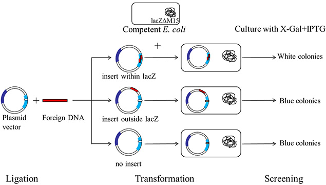
  • It is a rapid and efficient technique for the identification and recombinant bacteria.
  • It relies on the activity of β- galactosidase ( β- gal) an enzyme that occurs in E. coli which cleaves lactose into glucose and galactose.
Schematic representation of typical plasmid vector that can be used for blue-white screening.

Fig: Schematic representation of typical plasmid vector that can be used for blue-white screening.


LacZ gene & their Disruption:

  • It also called the heart of the Blue/white selection and is allows the selection of insert DNA fragment into the vector.

  • It has been engineered to contain multiple cloning sites (MCS), which is an oligonucleotide sequence with a series of different restriction endonuclease recognition sites arranged in tandem in the same reading frame as the lacZ gene itself.

  • The lacZ gene codes for the β- gal enzyme, which metabolizes the β- galactosidase bond into lactose. It will also cleave the galactosidase bond in an artificial substrate called X-gal (5-Bromo-4-chloro- 3-indoyl-beta-D-galactopyranoside), which can be added to bacterial growth media and has a blue color when cleaves by intact enzymes.


  • Most plasmid vectors carry a short segment of the lacZ gene that contains coding information for the first 146-amino acids of β- galactosidase. The host E.coli strains used are competent cells containing lacZΔM15 deletion mutation. 

  • When the plasmid vector is taken up by such cells, due to the α- complementation process, a functional β- galactosidase enzyme is produced. 

  • If the fragment of DNA is cloned (inserted) into multiple cloning sites (MCS), the lacZ gene will be disrupted, inactivating it, and the resulting β- galactosidase will no longer be able to cleave X-gal, resulting in white bacterial colonies rather than blue colonies.

How does Blue/white screening work:

For screening the clones containing recombinant DNA, a chromogenic substrate, X-gal is added to the agar plate. If β- galactosidase is produced, X-gal (substrate) is produced, X-gal is hydrolyzed to form 5-Bromo-4-chloro-indoxyl, which spontaneously dimerizes to produce an insoluble blue pigment called 5,5'-dibromo-4,4'-dichloro indigo. The colonies formed by non-recombinant cells, therefore appear blue in color while the recombinant ones appear white. The desired recombinant colonies can be easily picked and cultured.

Isopropyl β-D-1- thiogalactopyranoside (IPTG) is used along with X-gal for blue-white screening. IPTG (inducer) is a non-metabolizable analog of galactose that includes the expression of the lacZ gene.

A schematic representation of a typical blue-white screening procedure

Fig: A schematic representation of a typical blue-white screening procedure


Limitations of the Blue/white screening:

  • The lacZ gene in the vector may sometimes be non-functional and may not produce β- galactosidase. The resulting colony will not be recombinant but will appear white. 
  • Even if a small sequence of foreign DNA may be inserted into MCS and change the frame of the lacZ gene. This results in false-positive white colonies.
  • Small inserts within the reading frame of lacZ may produce ambiguous light blue colonies as β- galactosidase is only partially inactivated.

👉Previous Page:Telomere - CSIR NET/ICMR/ DBT (Life Sciences)

👉Next Page:Breast Cancer (BRCA1) gene binding partner- CSIR/ ICMR/ DBT (Life Sciences)

Share:

Saturday, May 15, 2021

Telomere - CSIR NET/ICMR/ DBT (Life Sciences)

Telomere

  • Telomeres are the terminal regions of the linear chromosome of eukaryotes (not is prokaryotes because of the circular chromosomes) composed of telomeric DNA and associated with specific proteins.

  • Telomeric DNA is associated with a large number of repetitive, strictly defined short nucleotide sequences or tandem repeats of 5'-TTAGGG-3' (vertebrates), TTTAGGG (terrestrial plants).


  • This sequence is usually repeated about 3,000 times and can reach up to 15,000 base pairs (bp) in length.
Telomere

Though telomeric proteins differ among different groups of organism they perform similar functions like;

  • telomere length regulations and protection against degradation.

For a long time, it was considered that telomeres did not code RNA molecule and thus proteins. Succeding, it was found that RNA is still transcribed from telomeres but it did not encode any protein. Further studies showed that this RNA plays an important role in;

  • telomere length regulations
  • chromatin reorganization during both development and cell differentiation.

Even though telomere does not encode protein. They also perform very important functions;

  • maintain the stability and functionality of the cellular genome.
  • help to organize each of our 46 (23 pairs) chromosomes in the nucleus of cells.
  • protects the end of the chromosome by forming a cap, much like the plastic tip on shoelaces. (If the telomeres were not there, the chromosome may end up sticking to other chromosomes).
  • protection of chromosomes from fusion with each other.
  • stabilization of broken chromosome ends.
  • attachment to the nuclear envelope.
  • influencing gene expression.
  • counting the quantity of cell division.
  • protection of the mRNA coding regions of the chromosome from the end replication problem.
  • participation in mitotic and meiotic chromosome segregation.

Telomeres dysfunction causes aging or cancer depending on the DNA damage response.

Factors that influence telomere function:

  • Telomerase
  • Telomeric chromatin
  • Shelterin complex


Telomerase:

Telomerase
  • It is a ribonucleoprotein called terminal transferase, that adds a species-dependent telomere repeat sequence to the 3' end of telomeres.

  • It is a two-partner enzyme, the reverse transcriptase catalytic subunit (TERT) and the RNA component (TERC), responsible for the maintenance of the length of telomeres by the addition of a G-rich repetitive sequence.

Telomeres need to be protected from a cell's DNA repair system because they have single-stranded overhangs, which "look like" damaged DNA. The overhang at the lagging strand end of the chromosome is due to incomplete end replication. The overhang at the leading strand end of the chromosome is generated by enzymes that cut away part of the DNA.


During each cell division cycle, telomeres shorten as a result of the incomplete replication of linear DNA molecules by conventional DNA polymerase. Telomerase compensates for telomere attrition through the addition of TTAGGG repeats by TERT onto the chromosome ends by using an associated RNA component as template TERC.


Shelterin Complex:

Shelterin Complex

  • Shelterin is a 6-subunit protein complex comprising TRF1, TRF2, POT1, TPP1, TIN2, RAP1, that associates with mammalian cells and allows cells to distinguish the natural ends of chromosomes from sites of DNA damage.

  • Shelterin binds telomeres through TRF1 and TRF2, which interact with the double-stranded (ds) telomeric DNA.

  • POT1 (POT1 & POT2 in the mouse) is associated with single-stranded (ss) telomeric DNA. 

  • POT1 is linked to TRF1 and TRF2 via an interaction between the POT1-binding protein TPP1 and TIN2, which binds both TRF1 and TRF2. 

  • RAP1 interacts solely with TRF2. It is dispensable for telomere capping but prevents telomere recombination and fragility. Thus RAP1 is not a telomere protective protein, in contrast to the rest of proteins. Hence, the role of RAP1 is telomerase regulation.

Function:

  • Shelterin function is crucial for telomere maintenance and genome integrity.
  • It protects telomeres from DNA damage signaling and DNA repair.
  • It promotes the semi-conservative replication of the telomeric DNA.
  • It regulates the telomerase-mediated maintenance of the telomeric DNA.


Telomere dysfunction and genome instability:

  • Genome instability is a prominent characteristic of most cancer types that have an essential role in tumorigenesis by accelerating the accumulation of genetic changes that are responsible for cancer cell evolution.

  • One of the important sources of genome instability is telomere shortening.

  • Telomere dysfunction can cause by a deficiency of telomerase and/or the shelterin proteins, either owing to the loss of the telomere protective structure, causes genome instability, and thereby affects tumorigenesis.

The molecular mechanism that related to telomere defects are:

  • Breakage fusion bridge cycles.
  • Defects in telomeric replication.
  • Susceptibility of telomeric DNA to genotoxic damage.
  • Cell cycle control and endoreduplication, replication of nuclear genome in the absence of mitosis, which leads to elevated nuclear gene content and polyploidy. E.g. Telomere shortening.


Share:

Thursday, May 6, 2021

Protein Folding- CSIR NET/ICMR/DBT (Life sciences)

Protein Folding

  • Protein folding is the physical process by which a polypeptide folds into its characteristics & functional three-dimensional 3D conformation.

  • The correct 3D structure is essential to protein function. Failure to fold into native structure produces inactive proteins.

  • A protein molecule folds spontaneously during/after biosynthesis. However, the process also depends on the nature of the solvent, the concentration of salts, the temperature, and the presence of molecular chaperones.

Protein Folding- CSIR NET/ICMR/DBT (Life sciences)


Experiment:

According to the experiment, which helps in the understanding of the protein folding was carried out by Biochemist Christian Anfinsen in 1960, that is the refolding of the protein ribonuclease A. 
  • Ribonuclease A isolated from the bovine pancreas is an enzyme that has a molecular weight (mw=13,700 Da) and it contains 124 amino acid residues and 4- disulfide linkages.

  • In the presence of a denaturant (Urea), and a reductant (reducing agent β-mercaptoethanol), Ribonuclease is denatured and the sulfide bonds are broken.

When the protein is allowed to renature by removing the denaturant and reductant (reducing agent), the protein regains its native confirmation, including 4 correctly paired disulfide bonds.

This finding provided the first evidence that;

  • However, when the reductant (reducing agent β-mercaptoethanol) is removed, while the denaturant (Urea) is still present, the disulfide bonds are formed again in protein but most of the disulfide bonds are formed between incorrect partners. This indicates that weak interactions are required for correct positioning of disulfide bonds and assumption of the native conformation.

Anfinsen's Experiment: Denaturation/Renaturation of Ribonuclease. Depending on the conditions for renaturation, we obtain either native ribonuclease or scrambled ribonuclease. 


Share:

Wednesday, January 13, 2021

Plant Tissue - NEET (Botany)

Plant Tissue-NEET (Botany)


Plant Tissue
:
A group of cells having essentially a common function and origin is called a tissue. The plant tissue is made up of a group of cells. These cells may be either similar or dissimilar in structure, function, and origin. Plant of higher-level shows this level of cellular organization.

Fig: Plant Tissue Flowchart

Plant tissues are broadly categorized into three tissue systems. This classification is based on parts of the plants that are present.

  • Epidermis Tissue: cells formed from the outermost surface of the leaves.

  • Vascular Tissue: involved in transporting fluid and nutrients internally.
  • Ground Tissue: involved in producing nutrients by photosynthesis and preserve nutrients.

Plant tissues are grouped as two types based on their ability to divide:

A. Meristematic Tissue. 
B. Permanent Tissue.


A. MERISTEMATIC TISSUE:

  • It is a group of young cells, which consists of continually dividing cells and helps in the increase of length and width of the plant.
  • These are living cells with the ability to divide into the regions where they are present.
  • These are polyhedral or isodiametric in shape without intercellular spaces.
  • The cell wall is thin, elastic, mainly composed of cellulose.
  • Protoplasm is dense with a distinct nucleus at the center and vacuoles if present, are very small.
  • Cells show a high rate of metabolism.
  • It is involved in the movement of water and nutrition within the plants.
  • These tissues are responsible for both the primary and secondary growth of the plant.
  • It is the outermost tissue, functions by providing protection from mechanical injury.
  • It gives rise to the epidermis layer, cortex, endodermis, ground tissue, and vascular tissue.

Classification of Meristem:

Following criteria are used for the classification of meristems viz. origin, function, and position as follows:

A. Origin:

Primordial Meristem or Promeristem

  • It is also called an embryonic meristem.
  • It is usually occupying a very minute are at the tip of the root and shoot.

Primary Meristem

  • It originates from the primordial meristem and occurs in the plant body from the beginning, at the root and shoot apices.
  • Cells are dividing and different permanent tissues are produced from primary meristems.

Secondary Meristem

  • It develops from living permanent tissues during later stages of plant growth; hence are called secondary meristems.
  • This tissue occurs in the mature regions of the root and shoot of many plants. 

B. Position:

Apical Meristem:

  • It is produced from promeristem and forms a growing point of apices of roots, shoots, and their lateral branches.
  • It brings about an increase in the length of the plant body and is called apical initials.
  • Shoot apical meristem is terminal in position whereas in root it is subterminal i.e. located below the root cap.
  • Intercalary meristematic tissue is present in the top or base area of the node.
  • Their activity is mainly seen in monocots.
  • They are short-lived.

Lateral Meristem:

  • It is present along the sides of a central axis of organs.
  • It takes part in increasing the girth of stem or root, eg. intrafascicular cambium.
  • It is found in vascular bundles of gymnosperms and dicot angiosperms.


C. Functions:

  • The young growing root of the plant has Protoderm that forms a protective covering like epidermis around the various organs.
  • Meristem called Procambium is involved in developing primary vascular tissue while the other structures like cortex, endodermis, pericycle medullary rays, pith are formed from the region of the ground meristem. These three groups of meristem are based on the functions.
B. PERMANENT TISSUE:
  • This is a group of cells that have lost the capacity of division and acquired permanent size, shape, and functions.
  • They are offer elasticity, flexibility, and strength to the plant.

Depending upon the types of cells, there are two types as simple and complex permanent tissues.


1. SIMPLE PERMANENT TISSUE:

  • These are made up of only one type of cells carrying similar functions.
  • This tissue is either living or dead.
  • The types of simple permanent tissues: Parenchyma, Collenchyma, Sclerenchyma.

A. Parenchyma:

  • Cells in this tissue are thin-walled, isodiametric, round, oval to polygonal, or elongated in shape.
  • The cell wall is composed of cellulose.
  • The cells are living with a prominent nucleus and cytoplasm with large vacuoles.
  • This is less specialized permanent tissue.
  • The parenchyma has distinct intercellular spaces. Sometimes, cells may show a compact arrangement.
  • The cytoplasm of adjacent cells is interconnected through plasmodesmata and thus forms a continuous tissue.
  • These cells are distributed in all the parts of the plant body viz. epidermis, cortex, pericycle, pith, mesophyll cells, endosperm, xylem, and phloem.

Functions:

  • The cells stores food, water, help in gaseous exchange, increase buoyancy, perform photosynthesis, and different functions in the plant body.
  • Differentiation in parenchyma cells develops vascular cambium and cork cambium at the time of secondary growth.
  • Some parenchymatous cells perform as storage chambers for starch in vegetables and fruits.


Fig: Parenchyma tissue


B. Collenchyma:
  • It is a simple permanent tissue made up of living cells.
  • The cell wall is cellulosic but shows uneven deposition of cellulose and pectin especially at corners.
  • The cell wall may show the presence of pits. 
  • The cells are similar to parenchyma containing cytoplasm, nucleus, and vacuoles but small in size and without intercellular gaps. Thus appears to be compactly packed.
  • Shape: either circular, oval, or angular in the transverse section.
  • Collenchyma is usually absent in monocots and roots of the dicot plant.

Functions:

  • Collenchyma is living mechanical tissue and serves different functions in plants.
  • It gives mechanical strength to young stems and parts like the petiole of the leaf.
  • It allows bending and pulling action in plant parts and also prevents tearing of leaf.
  • Growth of organs and elongation.

Fig: Collenchyma tissue


C. Sclerenchyma:
  • It is simple permanent tissue made up of compactly arranged thick-walled dead cells.
  • The cells are living at the time of production but at maturity they become dead.
  • As cells are devoid of cytoplasm their thickened walls are due to uniform deposition of lignin.
  • The cells remain interconnected through several pits.

It is of two types: 1.) Fibers & 2.) Sclerids.

1.) Fibers

  • It is a thread-like, elongated, and narrow structures with tapering and interlocking end walls.
  • These are mostly in bundles, pits are narrow, unbranched, and oblique.
  • They provide mechanical strength.

2.) Sclerids:

  • It is usually broad, with blunt end walls.
  • These occur singly or in loose groups and their pits are deep branched and straight.
  • These are developed due to the secondary thickening of parenchyma cells and provide stiffness only.

Functions:

  • This tissue functions as the main mechanical tissue.
  • It permits bending, shearing, and pulling.
  • It gives rigidity to leaves and prevents them from falling.
  • It also gives rigidity to epicarps and seeds.
  • Commercial fibers are also produced from sclerenchyma fibers. e.g. jute, flax, hemp.

2. COMPLEX PERMANENT TISSUE:
  • This tissue is heterogeneous comprising more than one type of cell and all function as a single unit.
  • This tissue is involved in conducting the sap and food from source to sink area.
  • This type of complex permanent tissue: Xylem, Phloem.
Fig: Complex Permanent Tissue


A. Xylem:
  • It is dead complex tissue.
  • The xylem also provides mechanical strength to the plant body.
  • Tracheids and Vessels conduct water and minerals. These are known as hadrome = the part of the xylem mestome that conducts water and nutrients.
  • In pteridophytes and gymnosperms, tracheids are conducting elements and vessels in angiosperms, Selaginella (Pteridophyte) and Gnetum (Gymnosperm) show the presence of vessels.

1.) Tracheids:

  • It is elongated, tubular, and dead cells.
  • The ends are oblique and tapering.
  • The cell walls are uniformly thickened and dignified. This provides mechanical strength.
  • It contributes 95% of the wood in Gymnosperms and 5% in Angiosperms.
  • The different types of thickening patterns are seen on their walls such as annular (in the form of rings), spiral (in the form of spring/helix), scalariform (ladder-like), pitted is the most advanced type (small circular area) which may be simple or bordered.

2.) Vessels:

  • It is longer than tracheids with perforated or dissolved ends and formed by the union of several vessels end to end.
  • These are involved in the conduction of water and minerals.
  • Their lumen is wider than tracheids and the thickening is due to lignin and similar to tracheids.
  • In monocot, vessels are rounded.
  • In Dicot angiosperm, vessels are angular.
  • The first formed xylem vessels (protoxylem) are small and have either annular or spiral thickening.
  • The later formed xylem vessels (metaxylem) have reticulate or pitted thickenings.
  • Endarch: When protoxylem is arranged towards pith and metaxylem towards the periphery. E.g. in the stem.

  • Exarch: When metaxylem is arranged towards pith and protoxylem towards the periphery. E.g. in the roots.

Fig: Tracheids & Vessels (Xylem tissue)


3.) Xylem Parenchyma (Only living tissue):
  • The cells are small associated with tracheids and vessels.
  • This is the only living tissue among the complex tissue.
  • The function is to store food (starch) and tannins.
  • It is involved in lateral or radial conduction of water or sap.

4.) Xylem Fibers:

  • The tissue is sclerenchymatous cells and serves mainly mechanical support.
  • These are called wood fibers.
  • Shape: elongated, narrow, spindle.
  • Cells are tapering at both ends and their walls are lignified.

B. Phloem (Bast):

  • This is a living tissue. It is called as bast.
  • Phloem is responsible for the conduction of organic food material from a source (generally leaf) to a sink (other plant parts).
  • It was named leptome by Haberlandt as similar to the xylem.
  • Based on origin, it is first formed (Proto) and lately formed (Meta) phloem.
  • It is composed of sieve cells, sieve tubes, companion cells, phloem parenchyma, and phloem fibers.

1.) Sieve tubes:

  • It is a long tubular conducting channel of phloem.
  • These are placed end to end with bulging at end walls.
  • The sieve tube has a sieve plate formed by septa with small pores.
  • The sieve plates connect the protoplast of the adjacent sieve tube cells.
  • The sieve tube cell is connected to a companion cell through phloem parenchyma by plasmodesmata.
  • Sieve cells are found in the lower plants like pteridophytes and gymnosperms.
  • The cells are narrow, elongated with tapering ends and a sieve are located laterally.

2.) Companion cells:

  • This is narrow elongated and living.
  • These cells are laterally associated with sieve tube elements.
  • The companion cells have dense cytoplasm and prominent nucleus.
  • The nucleus of the companion cell regulates the functions of the sieve tube cells through simple pits.
  • From an original point of view, the sieve tube cells and companion cells are derived from the same cell.
  • The death of the one results in the death of the other type.

Fig: Sieve tubes & Companion cells


3.) Phloem Parenchyma:
  • The cells are living, elongated found associated with sieve tube and companion cells.
  • The chief function is to store food, latex, resins, mucilage, etc.
  • The cells carry out lateral conduction of food material.

4.) Phloem Fibers (only Dead tissue):

  • These are the only dead tissue among the phloem. These are sclerenchymatous. 
  • It is generally absent in the primary phloem but present in the secondary phloem.
  • These cells are with lignified walls and provide mechanical support.
  • They are used in making ropes and rough clothes.


Share:

Unordered List

Search This Blog

Powered by Blogger.

About Me

My photo
Education marks proper humanity.

Text Widget

Featured Posts

Popular Posts